Full-Body MRI Screening: The Preventive Health Trend Taking Over
Early detection saves lives. Learn why high-resolution imaging is becoming the gold standard for identifying potential health issues years before symptoms appear.
OMICS Clinical Team · Medical review: Pending medical review
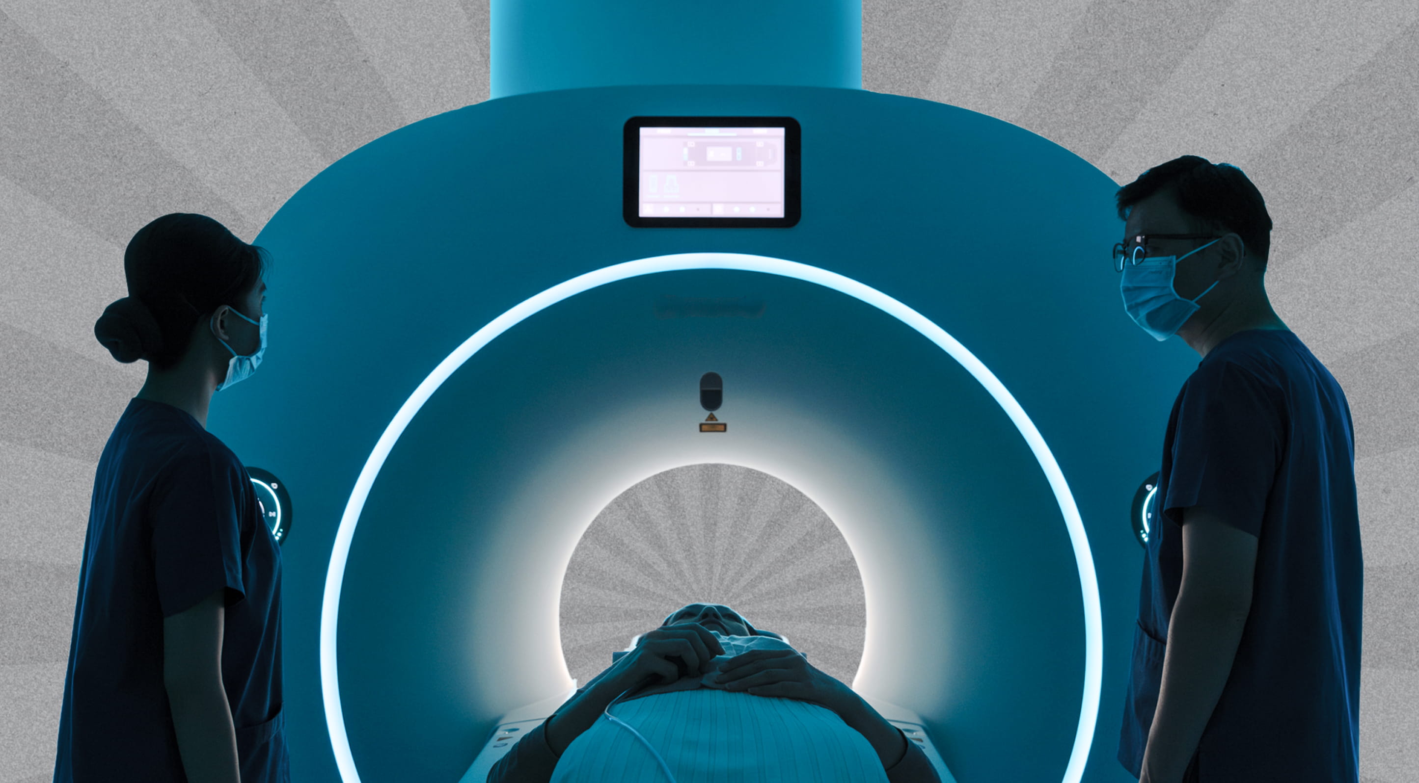
Full-body MRI screening has moved from a niche imaging service into one of the most discussed tools in preventative medicine. Adults who once relied on annual blood tests and a chest X-ray are now asking whether a single scan can offer something more meaningful: a structural look at the whole body, captured in roughly an hour, with no radiation. In Dubai, demand has grown alongside the city's broader shift toward precision diagnostics and longevity-focused care. The technology is genuinely useful, but it is also widely misunderstood. This guide explains what full-body MRI screening actually is, what it can and cannot detect, and where it fits within a serious preventative health strategy.
What Is Full-Body MRI Screening?
Full-body MRI screening is a non-invasive imaging examination that uses magnetic resonance technology to produce detailed images of the body's organs, soft tissues, blood vessels and skeletal structures in a single session. Unlike a targeted MRI focused on one anatomical region, a full-body protocol captures images from the brain through to the pelvis, and in many protocols extends down through the upper thighs.
The scan typically takes between 45 and 90 minutes depending on the protocol and the strength of the scanner. No ionising radiation is involved, no contrast agent is required for many screening protocols, and there is no recovery period. The output is a comprehensive set of cross-sectional images that a radiologist reviews for structural abnormalities, masses, vascular changes and signs of organ-specific disease.
The modern interest in full-body MRI as a screening tool, rather than a diagnostic one, is recent. Diagnostic MRI has been a cornerstone of clinical imaging for decades, but it has historically been ordered in response to symptoms or a specific clinical question. Screening MRI, by contrast, is performed in asymptomatic individuals to identify abnormalities before clinical signs appear. This is a meaningful shift in how the technology is used, and it sits at the centre of the current discussion about how preventative medicine should evolve.
Why Full-Body MRI Has Become a Preventative Health Trend
Several factors have converged to push full-body MRI screening into the mainstream of preventative care, particularly among adults focused on long-term health management.
The first is hardware progress. Higher field-strength scanners, faster sequences and improved image processing have made it feasible to scan the entire body in a clinically useful timeframe. What once required several hours can now be completed in roughly the duration of a film.
The second is a broader cultural movement toward proactive health. Adults in their forties, fifties and sixties are increasingly unwilling to wait for symptoms before investigating their health. The same population that has adopted continuous glucose monitoring, advanced lipid testing and biological age assessment is now asking whether structural imaging should be part of an annual review.
The third is the limitation of conventional screening. A standard annual check-up rarely includes imaging beyond a chest X-ray and abdominal ultrasound. Both have their place, but neither offers the resolution or coverage of MRI. Adults who have experienced a delayed diagnosis in family or social circles often arrive at preventative clinics asking for something more comprehensive.
The fourth is geography. In Dubai, where access to advanced medical technology is well established and where the executive population is unusually engaged with longevity medicine, full-body MRI screening has become one of the visible markers of a serious healthspan medicine approach. It is no longer an exotic offering. It is a recognised component of how precision diagnostics is delivered locally.
How Full-Body MRI Works
Magnetic resonance imaging relies on the behaviour of hydrogen atoms in water molecules within the body. When placed in a strong magnetic field, these atoms align in a predictable way. Pulses of radiofrequency energy then displace them briefly, and as they return to their original alignment they release signals. The scanner detects those signals and reconstructs them into detailed images.
The clinical advantage of MRI is its ability to differentiate between soft tissues with high contrast resolution. CT scans are excellent for bone and lung detail but are less able to distinguish between similar soft tissues without contrast. MRI can show subtle differences between healthy tissue, inflammation, fluid, fatty deposits and abnormal masses without exposing the patient to radiation.
A full-body screening protocol typically includes several sequence types. T1- and T2-weighted images provide complementary anatomical detail. Diffusion-weighted imaging is particularly useful for identifying lesions that behave differently from surrounding tissue, which is often the case with tumours and certain inflammatory processes. Some protocols add MR angiography to assess major blood vessels, or specialised cardiac sequences to evaluate heart structure.
The patient lies on a motorised table that moves through the scanner in stages. Coverage typically begins at the head and progresses through the neck, chest, abdomen, pelvis and upper thighs. Images are reviewed by a radiologist, ideally one with specific experience in whole-body screening interpretation, and findings are summarised in a structured report.
What Full-Body MRI Screening Can Detect
A well-conducted full-body MRI can identify a wide range of structural findings, some clinically significant and some incidental. Understanding the realistic detection profile is essential before considering a scan.
Possible Cancers and Masses
Full-body MRI can identify tumours and suspicious lesions in many organs, including the brain, liver, kidneys, pancreas, ovaries, prostate and lymphatic system. Diffusion-weighted sequences improve sensitivity for several cancer types, although detection capability varies by organ. MRI is generally less sensitive than dedicated low-dose CT for early lung cancer and is not a replacement for established cancer-specific screening such as mammography or colonoscopy. It is most useful as a complement to, not a substitute for, recommended population screening programmes.
Vascular Abnormalities
Aneurysms in the brain, aorta and other major arteries can often be identified on whole-body MRI, particularly when MR angiography sequences are included. Aortic dilatation, vascular malformations and certain congenital anomalies may be detected before they produce symptoms.
Organ-Specific Disease
Structural changes in the liver, kidneys, pancreas and adrenal glands can be visualised. Fatty liver, cysts, hemangiomas, adenomas and other benign or potentially significant findings are commonly identified. The scan can also detect renal abnormalities, biliary tree variations and certain endocrine masses.
Musculoskeletal and Spinal Findings
Disc disease, spinal cord abnormalities, joint pathology and bone marrow changes are visible on full-body protocols. While these findings are often incidental, they can occasionally be clinically meaningful, particularly when they correspond with reported symptoms or an existing clinical concern.
Neurological Findings
Brain MRI sequences within a whole-body protocol can identify structural lesions, vascular changes, signs of small vessel disease and incidental findings such as meningiomas. Image quality for the brain is generally adequate for screening but does not replace a dedicated neurological MRI when clinical concern is high.
What Full-Body MRI Cannot Reliably Do
Equally important is understanding the limitations. The technology is powerful, but it is not a universal detector of disease, and several common misconceptions need addressing.
It does not assess function. MRI shows structure, not how an organ is performing. A scan cannot evaluate kidney function, liver enzymes, hormonal output, immune activity or metabolic regulation. These remain the domain of laboratory testing.
It does not replace cancer-specific screening. For certain cancers, dedicated screening modalities are more sensitive at detecting early disease. Mammography for breast cancer, colonoscopy for colorectal cancer and low-dose CT for lung cancer in eligible smokers each have established evidence bases that whole-body MRI does not displace.
It does not evaluate the coronary arteries in detail. Cardiac MRI provides important structural information about the heart muscle, chambers and valves, but it is not the optimal modality for assessing coronary artery plaque burden. Coronary artery calcium scoring with low-dose CT remains the more direct tool for that purpose.
It does not detect everything that matters. Microscopic disease, early metabolic changes, inflammatory processes that have not yet produced structural change, and many cancers below a certain size threshold will not be visible on any imaging study.
It produces incidental findings. This is one of the most important caveats. A whole-body scan in a typical adult often identifies findings that are not clinically significant. Cysts, benign lesions, mild structural variations and age-related changes are common. Each requires careful interpretation, and in some cases follow-up imaging or further investigation, which can introduce anxiety, additional cost and occasionally unnecessary procedures.
The Incidentaloma Problem
Any honest discussion of full-body MRI screening must address the question of incidental findings, often called incidentalomas. Published studies of whole-body MRI in asymptomatic adults consistently show that incidental findings are common, with reported rates varying widely depending on the population studied and how findings are categorised.
Most incidental findings are benign or of uncertain clinical significance. The challenge is that determining benignity often requires further investigation: repeat imaging, blood tests, specialist consultation, and occasionally biopsy. For each genuinely important finding identified early, there are typically several findings that require follow-up but ultimately turn out to be inconsequential.
This is not an argument against full-body MRI. It is an argument for ensuring that the scan is performed and interpreted within a clinical framework that can manage the findings appropriately. A scan ordered without medical oversight, interpreted in isolation, and delivered as a written report without expert discussion is a recipe for either undue alarm or false reassurance. A scan ordered as part of a structured preventative diagnostics programme, interpreted by a radiologist with screening experience, and discussed with a physician who knows the patient is a fundamentally different experience.
The clinical value of full-body MRI screening is not the scan itself. It is the integration of the scan into a coherent assessment of risk and a considered plan for follow-up.
Who Is Best Suited to Full-Body MRI Screening?
Full-body MRI screening is not equally appropriate for everyone. Several factors influence whether it is likely to provide meaningful clinical value.
Adults over 40 are generally the population in which structural findings of relevance become more frequent. The yield of clinically actionable findings rises with age, although younger adults with specific risk factors may also benefit.
Individuals with a meaningful family history of cancer, vascular disease or other conditions where structural imaging adds information may find a baseline scan particularly useful. Family history does not determine personal outcome, but it shifts the prior probability of relevant findings and can influence how findings are interpreted.
Adults engaged in proactive health management who are already monitoring biomarkers, biological age and metabolic indicators may find that full-body imaging completes a structural layer that laboratory testing alone cannot provide.
People with vague or persistent symptoms that have not been explained by standard investigations sometimes benefit from a comprehensive scan, although in this scenario the scan is closer to diagnostic imaging than to pure screening.
The scan is generally less appropriate for individuals with severe claustrophobia (although open and wide-bore scanners exist), those with certain implanted medical devices that are not MRI-compatible, and those whose health priorities would be better served by addressing established risk factors before adding imaging.
It is also worth being candid that for younger adults with no family history, no symptoms and no significant risk factors, the likelihood of a full-body MRI changing clinical management is lower. The decision to scan should be informed by clinical context rather than enthusiasm alone.
Full-Body MRI Screening in Dubai
Dubai has developed one of the more comprehensive medical imaging infrastructures in the region. Several hospitals and specialist clinics offer full-body MRI screening, and the protocols available locally are typically aligned with international standards. For residents and visitors who want a single, comprehensive structural assessment, access is rarely the limiting factor.
What distinguishes how the scan is delivered is the clinical context surrounding it. In a transactional model, full-body MRI is offered as a standalone product: book a scan, attend the appointment, receive a report. This model has its uses but tends to amplify the limitations of screening imaging without providing the framework needed to act on findings constructively.
A precision diagnostics model integrates the scan into a broader assessment. Laboratory testing, biomarker profiling, biological age measurement and structural imaging are combined into a single clinical picture. Findings are interpreted in relation to one another rather than in isolation. A small lesion identified on MRI is considered alongside inflammatory markers, family history, hormonal status and metabolic indicators. The result is a more informative and more actionable assessment than the imaging alone could provide.
Dubai's executive population, in particular, has gravitated toward this integrated model. The reason is straightforward. A scan that produces a list of findings is useful only if those findings are translated into a plan, and the plan is only as good as the clinical thinking behind it. The infrastructure required for that level of integration, advanced imaging, multi-omic laboratory capability and physician-led interpretation, is now reliably available in the city.
Full-Body MRI Compared to Other Screening Approaches
Understanding where full-body MRI fits requires comparing it to alternative imaging and screening modalities.
Standard annual check-up imaging typically includes a chest X-ray and an abdominal ultrasound. These are useful for specific purposes but offer limited resolution and limited coverage. They cannot replicate the structural detail of MRI.
Low-dose CT screening has an established role in lung cancer screening for eligible high-risk adults. CT also provides excellent visualisation of bone and pulmonary structures. Its limitations are radiation exposure, limited soft-tissue contrast compared with MRI, and the need for contrast agents in many protocols.
Coronary artery calcium scoring, performed by low-dose CT, is one of the strongest predictors of cardiovascular events. It is not a substitute for full-body MRI and addresses a different question: how much calcified plaque is present in the coronary arteries.
PET-CT is sometimes used in advanced screening protocols, particularly for cancer surveillance in higher-risk populations. It involves both radiation and a radioactive tracer and is generally used in defined clinical scenarios rather than routine screening.
Ultrasound remains a valuable, radiation-free tool for specific organ assessment, including the carotid arteries, thyroid, abdomen and pelvis. Its main limitation is operator dependence and limited tissue depth in some applications.
Standard MRI of a single region is performed when there is a specific clinical question, such as evaluating a known knee injury or investigating a suspected brain abnormality. Whole-body MRI broadens this to a screening application.
In practice, these modalities are complementary rather than competing. A serious preventative diagnostics programme draws on several of them in combination, selecting the right tool for each clinical question rather than relying on any single technology.
How Often Should Full-Body MRI Screening Be Repeated?
There is no single internationally agreed schedule for repeat full-body MRI screening. The appropriate interval depends on baseline findings, risk profile and clinical judgement.
For an asymptomatic adult with a clean baseline scan and no significant risk factors, a repeat interval of two to three years is often considered reasonable. For individuals with specific risk factors, family history of relevant disease or findings on baseline imaging that warrant monitoring, shorter intervals may be appropriate.
What matters more than the precise interval is that the decision is made within a clinical relationship rather than as a calendar reminder. A repeat scan in someone whose risk profile has not changed and whose baseline was unremarkable adds limited value. A repeat scan that follows up on a specific finding, or that is timed to a meaningful change in clinical context, adds substantially more.
Frequently Asked Questions
Is full-body MRI screening safe?
Full-body MRI uses no ionising radiation, which is one of its principal advantages. The procedure is generally considered safe for most adults. Individuals with certain implanted devices, such as some pacemakers, cochlear implants or specific metallic implants, may not be eligible, and a screening questionnaire is completed before the scan. Severe claustrophobia can be a practical limitation, although wide-bore and open scanners reduce this issue for many patients.
How long does a full-body MRI screening take?
A typical full-body screening protocol takes 45 to 90 minutes depending on the scanner, the sequences performed and any additional regions assessed. The patient lies still during the scan, and most modern scanners include features such as ventilation, lighting and audio that improve comfort during the examination.
Will full-body MRI detect cancer?
Full-body MRI can identify many structural abnormalities, including some early-stage cancers, particularly in solid organs such as the liver, kidneys, pancreas and brain. It is not a comprehensive cancer screening tool. Certain cancers, including some early lung cancers and microscopic disease, may not be visible on MRI. The scan is best understood as one component of a broader cancer awareness strategy that also includes recommended population screening, family history assessment and clinical evaluation.
What is the difference between full-body MRI screening and a standard annual check-up?
A standard annual check-up usually focuses on blood tests, basic physical assessment, an ECG, a chest X-ray and an abdominal ultrasound. Full-body MRI screening adds detailed structural imaging of the brain, neck, chest, abdomen, pelvis and other regions in a single examination. The two are complementary. Annual blood-based assessment provides functional and biochemical information; full-body MRI provides structural information that the standard check-up does not capture.
How much does full-body MRI screening cost in Dubai?
Pricing varies significantly between providers, scanner specifications and protocol depth. Costs typically depend on whether the scan is offered as a standalone service or as part of an integrated preventative diagnostics programme, and on whether additional sequences such as MR angiography or cardiac assessment are included. Insurance coverage is often limited for screening MRI, although policies vary, and individuals are advised to confirm coverage in advance with their provider.
Can full-body MRI replace other recommended screening tests?
No. Full-body MRI does not replace established screening programmes such as mammography, cervical screening, colonoscopy or eligible lung cancer screening. These programmes are based on substantial evidence and remain the recommended approach for the conditions they target. Full-body MRI complements them by providing structural assessment of regions and tissues that population screening does not cover.
What happens if the scan finds something unexpected?
Incidental findings are common in screening imaging. Most are benign or of uncertain clinical significance. When a finding is identified, the next step depends on its nature and the patient's overall clinical context. Options can include repeat imaging at a defined interval, additional targeted imaging, laboratory testing or specialist consultation. Findings are most usefully interpreted within an integrated clinical framework, ideally with a physician who can place the finding in the context of the patient's overall risk profile.
A Considered Place Within Preventative Diagnostics
Full-body MRI screening is a meaningful addition to modern preventative medicine, but it is not a shortcut. Its value is at its highest when it is used as part of a structured assessment that includes laboratory testing, biomarker analysis, biological age measurement and clinical interpretation. Used in isolation, it can produce more anxiety than insight. Used in context, it provides a structural layer of information that can genuinely support long-term health planning.
For adults in Dubai who are already engaged with precision diagnostics, full-body MRI screening represents a logical extension of an evidence-led approach. It is not a trend in the dismissive sense of the word. It is a technology with real clinical utility, becoming more accessible at the same moment that interest in proactive health management is reaching a critical mass. The thoughtful question is not whether full-body MRI screening is worth doing in principle, but whether and how it fits within a coherent personal health strategy that is informed by clinical judgement rather than enthusiasm alone.